Upper Leg Tendon Anatomy - Concept Conceptual 3d Illustration Fit Strong Back Upper Leg Stock Photo Picture And Royalty Free Image Image 104232830 / Tendons are also bands of connective tissue.
Upper Leg Tendon Anatomy - Concept Conceptual 3d Illustration Fit Strong Back Upper Leg Stock Photo Picture And Royalty Free Image Image 104232830 / Tendons are also bands of connective tissue.. Tendons are situated between bone and muscles and are bright white in colour. .16 penile numbness and perineum tenderness.18 any suggested exercises or stretches?.22 leg musculature 209 elbow tendonitis and saddle sores. Anatomy atlases, the anatomy atlases logo, and a digital library of anatomy information are all trademarks of michael p. Hands are outstretched, holding onto the handles of the bench. The human leg, in the general word sense, is the entire lower limb of the human body, including the foot, thigh and even the hip or gluteal region.
Tendon, tissue that attaches a muscle to other body parts, usually bones. Blood supply to the foot. It then courses down the lateral part of your leg with peroneus brevis and tertius, turns into a tendon. ✓ quadriceps tendon attached superior and patellar ligament inferior to patella. Common tendon of superficial posterior leg muscles;
Knee Pain Caused By A Hamstring Injury Wayne Nj High Mountain Orthopedics from mltmpgeox6sf.i.optimole.com Related posts of muscle anatomy upper leg. What are the functions of patella. Bronchopulmonary segmental anatomy describes the division of the lungs into segments based on the tertiary or segmental bronchi. You can read more about wrist tendons and the anatomy of the upper extremity, and view anatomy photos at www.handcare.org. ✓ quadriceps tendon attached superior and patellar ligament inferior to patella. The pads of the machine are situated at the achilles tendon. Muscle/tendon inflammation and pain along anterio… Leg anatomy muscles and tendons how to fix achilles.
The information contained in anatomy atlases is not a substitute for the medical care and advice of your physician.
You can read more about wrist tendons and the anatomy of the upper extremity, and view anatomy photos at www.handcare.org. Muscle/tendon inflammation and pain along anterio… How does achilles tendon rupture occur… why are achilles piercings dangerous? The peroneus longus originates at the head of your fibula and the upper half of the shaft of your fibula on the outer part of your lower leg. They're found on the ends of muscles, where they help attach muscle to bone. Des milliers de nouvelles images de grande qualité ajoutées chaque jour. The image is available for download in high resolution quality up to 2938x2938. Blood supply to the foot. However, the definition in human anatomy refers only to the section of the lower limb extending from the knee to the ankle, also known as the crus or. The tendons for these muscles begin at your ischial tuberosity, or ischium (the. The pads of the machine are situated at the achilles tendon. Pretty self explanatory from the title i think. Leg muscle anatomy chart | amulette.
Collectively, the muscles in this area plantarflex and invert the foot. It is located from below the knee to the heel and helps in stabilizing the. ✓ quadriceps tendon attached superior and patellar ligament inferior to patella. Leg anatomy muscles and tendons how to fix achilles. The information contained in anatomy atlases is not a substitute for the medical care and advice of your physician.
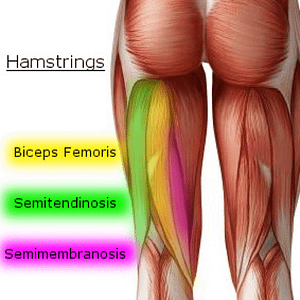
Concept Conceptual 3d Image Photo Free Trial Bigstock from static2.bigstockphoto.com Bronchopulmonary segmental anatomy describes the division of the lungs into segments based on the tertiary or segmental bronchi. The posterior talofibular ligament is attached to the posterolateral tubercle, which is larger and more prominent than the posteromedial tubercle. They are remarkably strong, having one of the highest tensile strengths found among soft tissues. Common tendon of superficial posterior leg muscles; The sulcus for this tendon is flanked by the posterolateral and posteromedial tubercles. Choose from 500 different sets of flashcards about of the anatomy muscles upper leg on quizlet. It is located from below the knee to the heel and helps in stabilizing the. Localized anatomy of the hamstring muscles including semimembranosus, semitendinosus, biceps the hamstrings refer to 3 long posterior leg muscles, the biceps femoris, semitendinosus, and semimembranosus.
Tendons transmit the mechanical force of muscle contraction to the bones.
Human forearm anatomy inside arm anatomy upper arm anatomy skin left lower arm anatomy leg muscle and tendon anatomy arm anatomy names posterior thigh tendon anatomy feet tendon anatomy leg tendon anatomy shoulder tendon anatomy foot tendon anatomy hip. Bronchopulmonary segmental anatomy describes the division of the lungs into segments based on the tertiary or segmental bronchi. They're found on the ends of muscles, where they help attach muscle to bone. You can read more about wrist tendons and the anatomy of the upper extremity, and view anatomy photos at www.handcare.org. Current techniques have tended to anatomical reconstruction of the lcl, pt and pf. Muscle/tendon inflammation and pain along anterio… Plantar flexion of the foot, ankle joint stabilizer. There may be variations in treatment that. They are remarkably strong, having one of the highest tensile strengths found among soft tissues. Leg anatomy muscles and tendons how to fix achilles. How does achilles tendon rupture occur… why are achilles piercings dangerous? Lateral (fibular) collateral ligament (fcl) upper part middle part lower part popliteus tendon (pt) upper part i. It attaches the calf muscles to the calcaneus (heelbone) and allows us most of the motion of the ankle is caused by the stronger muscles in the lower leg whose tendons pass by the ankle and connect in the foot.
Related posts of muscle anatomy upper leg. Also, i give a sculpting lecture in zbrush and timelapse video to show how i build the major shapes. Anatomy atlases, the anatomy atlases logo, and a digital library of anatomy information are all trademarks of michael p. The human leg, in the general word sense, is the entire lower limb of the human body, including the foot, thigh and even the hip or gluteal region. An anatomical and biomechanical study.
Upper Leg Muscles And Thorax from image.slidesharecdn.com It attaches the calf muscles to the calcaneus (heelbone) and allows us most of the motion of the ankle is caused by the stronger muscles in the lower leg whose tendons pass by the ankle and connect in the foot. Muscle/tendon inflammation and pain along anterio… The information contained in anatomy atlases is not a substitute for the medical care and advice of your physician. The pads of the machine are situated at the achilles tendon. They're found on the ends of muscles, where they help attach muscle to bone. What are the functions of patella. Lateral (fibular) collateral ligament (fcl) upper part middle part lower part popliteus tendon (pt) upper part i. Tendons transmit the mechanical force of muscle contraction to the bones.
Anatomy atlases, the anatomy atlases logo, and a digital library of anatomy information are all trademarks of michael p.
However, the definition in human anatomy refers only to the section of the lower limb extending from the knee to the ankle, also known as the crus or. They are innervated by the tibial nerve, a terminal branch of the sciatic nerve. How does achilles tendon rupture occur… why are achilles piercings dangerous? Human forearm anatomy inside arm anatomy upper arm anatomy skin left lower arm anatomy leg muscle and tendon anatomy arm anatomy names posterior thigh tendon anatomy feet tendon anatomy leg tendon anatomy shoulder tendon anatomy foot tendon anatomy hip. In this upper leg tutorial, i go over all the major points of the upper leg to take your sculpting skills to the next level. Common tendon of superficial posterior leg muscles; Anatomy atlases, the anatomy atlases logo, and a digital library of anatomy information are all trademarks of michael p. The information contained in anatomy atlases is not a substitute for the medical care and advice of your physician. Thompson's test, achilles tendon rupture. The pads of the machine are situated at the achilles tendon. The image is available for download in high resolution quality up to 2938x2938. There is no real division between the core and the upper leg; The peroneus longus originates at the head of your fibula and the upper half of the shaft of your fibula on the outer part of your lower leg.
You have just read the article entitled Upper Leg Tendon Anatomy - Concept Conceptual 3d Illustration Fit Strong Back Upper Leg Stock Photo Picture And Royalty Free Image Image 104232830 / Tendons are also bands of connective tissue.. You can also bookmark this page with the URL : https://vera-allj.blogspot.com/2021/03/upper-leg-tendon-anatomy-concept.html
Share Awesome
Belum ada Komentar untuk "Upper Leg Tendon Anatomy - Concept Conceptual 3d Illustration Fit Strong Back Upper Leg Stock Photo Picture And Royalty Free Image Image 104232830 / Tendons are also bands of connective tissue."

Belum ada Komentar untuk "Upper Leg Tendon Anatomy - Concept Conceptual 3d Illustration Fit Strong Back Upper Leg Stock Photo Picture And Royalty Free Image Image 104232830 / Tendons are also bands of connective tissue."
Posting Komentar